Ultimate EKG Cheat Sheet to Master: how to interpret heart rhythms and arrhythmia fast in 2025

If you’re looking for a quick, clear, and effective way to learn how to interpret heart rhythms and arrhythmia fast in 2025, this ultimate EKG cheat sheet is exactly what you need. Designed for nurses, medical students, and healthcare professionals, this guide simplifies the process of reading and understanding EKGs. It covers all essential heart arrhythmias in a concise, easy-to-follow format—making it the perfect resource to master how to interpret heart rhythms and arrhythmia fast in 2025, whether you’re preparing for an exam or working in a fast-paced clinical setting.
Electrocardiography (EKG) is one of the most powerful and widely used diagnostic tools for assessing cardiac health. By placing electrodes on the skin, an EKG detects the heart’s electrical impulses and converts them into waveforms that reveal vital information about heart rhythm. These patterns help medical professionals determine if a patient’s heartbeat is normal or irregular. When these electrical signals become disordered, they result in arrhythmias—abnormal heart rhythms caused by disruptions in the conduction system. This cheat sheet gives you a fast-track method to recognize, interpret, and respond to these changes with confidence.
GRAPH INTERPRETATION https://lifesavercpr.net/wp-content/uploads/2012/05/ECG-Rythum-Study-Guide.pdf?
Interpreting EKGs with Confidence: How to Interpret Heart Rhythms and Arrhythmia Fast 2025
Have you ever wondered how nurses and doctors can glance at an EKG strip and instantly identify life-threatening arrhythmias? Mastering how to interpret heart rhythms and arrhythmia fast in 2025 may seem daunting, but with the right foundation and tools—like this ultimate cheat sheet—you can build the skills to interpret EKGs accurately and efficiently.
Learning how to interpret heart rhythms and arrhythmia fast in 2025 requires a solid grasp of cardiac conduction, waveform patterns, and the mechanisms behind common and complex arrhythmias. With consistent practice and the help of visual aids like rhythm strips, you’ll begin to recognize key differences—such as atrial fibrillation vs. atrial tachycardia—at a glance. Though reading EKGs may seem like decoding a foreign language at first, with time and exposure, you’ll become fluent in interpreting even the most irregular rhythms.
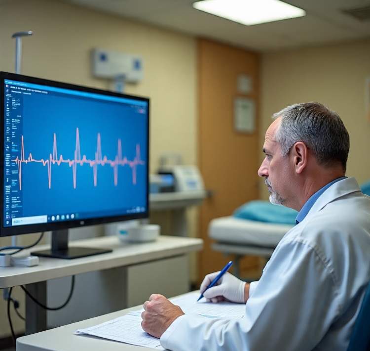
Example: Sinus Tachycardia
Understanding the characteristics of common rhythms like sinus tachycardia is a vital step in learning how to interpret heart rhythms and arrhythmia fast in 2025.
-
Definition: Sinus tachycardia is a heart rhythm with a rate greater than 100 beats per minute that originates from the sinus node.
-
Rate: 100–180 bpm
-
P Waves: Present and precede every QRS complex
-
PR Interval: Normal
-
QRS Complex: Normal
-
Conduction: Normal
-
Rhythm: Regular
Causes of sinus tachycardia may include physical exertion, stress, anxiety, fever, anemia, hypovolemia, drugs, heart failure, or shock. While sinus tachycardia is often asymptomatic, treatment is focused on addressing the underlying cause. In some cases, interventions such as carotid massage or beta-blockers may be used to reduce heart rate.
This rhythm is a perfect example to practice on as you learn how to interpret heart rhythms and arrhythmia fast in 2025. Identifying sinus tachycardia correctly can help prevent misdiagnosis and guide appropriate interventions.
Sinus Bradycardia
As you continue learning how to interpret heart rhythms and arrhythmia fast in 2025, understanding sinus bradycardia is essential. This rhythm originates from the sinus node and presents with a heart rate less than 60 beats per minute. Though it can be a normal variant in athletes, it may also signal underlying issues.
EKG Characteristics:
-
Rate: Less than 60 bpm
-
P waves precede each QRS complex
-
PR interval: Normal
-
QRS complex: Normal
-
Conduction: Normal
-
Rhythm: Regular
Causes: Drugs (like beta-blockers), vagal stimulation, hypothyroidism, hypothermia, or myocardial infarction.
Symptoms: Often asymptomatic but may include dizziness, fatigue, or syncope.
Management: Address the underlying cause; atropine sulfate may be used for symptomatic bradycardia.
Premature Atrial Contraction (PAC)
When studying how to interpret heart rhythms and arrhythmia fast in 2025, you’ll come across PACs—extra beats that originate from the atria. Though not true rhythms, they can signal atrial irritability.
EKG Characteristics:
-
Early and abnormal-looking P waves (different from normal sinus P waves)
-
QRS complex follows the premature P wave
-
P waves may be hidden in preceding T waves
Causes: Coronary artery disease, heart failure, COPD, hypoxia, electrolyte imbalance, or valvular disease.
Management: Typically benign; may use antiarrhythmic medications like quinidine or perform carotid sinus massage if symptomatic.
Atrial Flutter
To truly master how to interpret heart rhythms and arrhythmia fast in 2025, spotting the sawtooth pattern of atrial flutter is crucial. This rhythm originates in the atria and is often regular, but rapid.
EKG Characteristics:
-
Atrial rate: 250–400 bpm
-
Sawtooth-shaped flutter (P) waves
-
QRS: Uniform but may have irregular ventricular response
-
Rhythm: Atrial regular; ventricular may be irregular
Causes: Heart failure, valve disease (tricuspid or mitral), PE, cor pulmonale, inferior wall MI, carditis, or digoxin toxicity.
Management: If unstable and HR >150 bpm, immediate cardioversion. If stable, medications like beta-blockers, calcium channel blockers, or anticoagulants may be indicated.
Atrial Fibrillation (AFib)
A hallmark rhythm in the journey of learning how to interpret heart rhythms and arrhythmia fast in 2025, atrial fibrillation is a chaotic, disorganized rhythm of the atria.
EKG Characteristics:
-
Atrial rate: 350–600 bpm
-
Ventricular rate: 120–200 bpm
-
No discernible P waves; irregular baseline
-
Irregularly irregular rhythm
-
QRS: Normal
Causes: Atherosclerosis, HF, congenital heart disease, COPD, hyperthyroidism or hypothyroidism.
Symptoms: Palpitations, dyspnea, pulmonary edema.
Management: Control ventricular rate, reduce atrial irritability, address underlying cause. Anticoagulation may be required.
GRAPH INTERPRETATIONhttps://lifesavercpr.net/wp-content/uploads/2012/05/ECG-Rythum-Study-Guide.pdf?
Premature Junctional Contractions (PJC)
Another arrhythmia you’ll need to identify as you learn how to interpret heart rhythms and arrhythmia fast in 2025 is the PJC, which results from early impulses originating near the AV junction.
EKG Characteristics:
-
Inverted P wave (may precede, follow, or be buried in QRS)
-
PR interval < 0.12 sec
-
QRS complex: Normal
-
Rhythm: Irregular
Causes: MI, ischemia, digoxin toxicity, excessive caffeine or stimulant use.
Management: Eliminate the underlying trigger, adjust medications like digoxin if necessary.
Atrioventricular (AV) Blocks
AV blocks are conduction delays or interruptions between the atria and ventricles. Understanding the different degrees is key when learning how to interpret heart rhythms and arrhythmia fast in 2025.
First-Degree AV Block
EKG Characteristics:
-
Rate: 60–100 bpm
-
PR interval > 0.20 sec (consistently prolonged)
-
QRS complex: Normal
-
Rhythm: Regular
Causes: Inferior MI, ischemia, electrolyte imbalance, digoxin toxicity.
Management: Usually asymptomatic; monitor or treat if symptomatic with atropine.
Second-Degree AV Block (Mobitz I – Wenckebach)
EKG Characteristics:
-
Atrial rhythm: Regular
-
Ventricular rhythm: Irregular
-
PR interval progressively lengthens until QRS is dropped
-
PR interval resets after dropped beat
Causes: Inferior MI, cardiac surgery, vagal tone, digoxin.
Management: Correct underlying cause; atropine or temporary pacing if symptomatic.
Second-Degree AV Block (Mobitz II)
EKG Characteristics:
-
Atrial rhythm: Regular
-
Ventricular rhythm: Regular or Irregular
-
QRS periodically dropped
-
P–P interval constant
Causes: Severe CAD, anterior MI, myocarditis, digoxin toxicity.
Management: Emergency treatment with atropine, epinephrine, dopamine. Pacemaker often required.
Third-Degree AV Block (Complete Heart Block)
EKG Characteristics:
-
Atrial and ventricular rhythms: Regular but independent
-
No relationship between P waves and QRS
-
QRS: May be wide and bizarre
Causes: Congenital conditions, MI, hypoxia, LEv’s or Lenegre’s disease, digoxin toxicity.
Management: Atropine, dopamine, epinephrine. Most cases require permanent pacemaker.
Premature Ventricular Contractions (PVCs)
When diving deep into how to interpret heart rhythms and arrhythmia fast in 2025, you’ll frequently encounter PVCs—early beats originating from the ventricles due to increased automaticity in ventricular muscle cells. While often benign, PVCs require attention when they appear frequently or in specific patterns.

EKG Characteristics:
-
Atrial rhythm: Regular
-
Ventricular rhythm: Irregular
-
QRS complex: Premature, wide (>0.14 sec), and distorted
-
Often followed by a full compensatory pause
-
May appear singly, in pairs, or triplets
Causes: Myocardial ischemia or infarction, electrolyte imbalances (hypokalemia, hypomagnesemia), drug toxicity (digoxin, tricyclics), stimulants (caffeine, nicotine), and stress.
Clinical Clues: Usually asymptomatic, but may present with palpitations, lightheadedness, or weakness.
Management: Identify and treat the underlying cause. Administer antiarrhythmics (e.g., lidocaine, amiodarone) if indicated, especially when PVCs are multifocal or frequent (>6 per minute), or fall on a T wave (“R-on-T” phenomenon).
Ventricular Tachycardia (VT)
A core rhythm to master as you study how to interpret heart rhythms and arrhythmia fast in 2025 is ventricular tachycardia. Defined as three or more consecutive PVCs, VT can be life-threatening and demands immediate recognition.
EKG Characteristics:
-
Rate: 100–250 bpm
-
P waves: Often absent or buried in QRS
-
QRS complex: Wide and bizarre
-
PR interval: Not measurable
-
Rhythm: Usually regular
-
T waves often deflected opposite to QRS direction
Causes: MI, coronary artery disease, aneurysm, mitral valve prolapse, electrolyte imbalances, and drug toxicity (digoxin, epinephrine).
Clinical Signs: Lightheadedness, hypotension, dyspnea, or even loss of consciousness.
Management:
-
With pulse and stable: Follow ACLS guidelines—administer amiodarone, consider synchronized cardioversion if ineffective.
-
Pulseless VT: Immediate CPR, defibrillation, epinephrine, or vasopressin as per ACLS protocol.
Ventricular Fibrillation (VFib)

VFib is one of the most dangerous rhythms you’ll learn to identify while mastering how to interpret heart rhythms and arrhythmia fast in 2025. This rhythm results in chaotic, ineffective ventricular activity—leading to cardiac arrest if not treated immediately.
EKG Characteristics:
-
Rate: Rapid and disorganized
-
Rhythm: Chaotic, no discernible P waves or QRS complexes
-
QRS: Wide, erratic, and irregular
Causes: Acute MI, untreated VT, electrolyte imbalances, hypothermia, electric shock, and drug toxicity (digoxin, quinidine).
Symptoms: Sudden collapse, absence of pulse, respiration, or BP; may lead to seizures or death.
Management: Initiate CPR immediately, defibrillate, and follow full ACLS protocol (including epinephrine or vasopressin, airway management, and antiarrhythmics like amiodarone or lidocaine).
Additional Arrhythmias to Know in 2025
In addition to the rhythms covered above, anyone serious about how to interpret heart rhythms and arrhythmia fast in 2025 must also recognize:
-
Atrial Tachycardia – Rapid rhythm originating in the atria; treat based on cause and hemodynamic stability.
-
Torsades de Pointes – A polymorphic VT associated with prolonged QT interval; treat with magnesium sulfate and pacing.
-
Supraventricular Tachycardia (SVT) – Narrow complex tachycardia with rapid onset/offset; vagal maneuvers or adenosine often effective.
-
ST Depression – A sign of myocardial ischemia or non-STEMI; monitor and initiate appropriate cardiac care.
-
Asystole – Absence of ventricular activity; initiate CPR and ACLS protocol immediately—defibrillation is not indicated.
Download Your Printable EKG Cheat Sheet
PDF SOURCEhttps://lifesavercpr.net/wp-content/uploads/2012/05/ECG-Rythum-Study-Guide.pdf?
For fast reference and better retention while learning how to interpret heart rhythms and arrhythmia fast in 2025, download our Ultimate EKG Interpretation Cheat Sheet:
-
Part 1 – Basics of EKG Interpretation
-
Part 2 – Rhythm Strip Analysis
-
Part 3 – Arrhythmias at a Glance (with causes, symptoms, and treatment)
Common Arrhythmias: Description, Causes, and Treatments
| Arrhythmia | Description | Causes | Treatment |
|---|---|---|---|
| Sinus Arrhythmia | Irregular atrial & ventricular rhythm; normal P waves. | Common in athletes, elderly; digoxin toxicity; inferior wall MI. | Usually none. Atropine if HR < 40 bpm. |
| Sinus Tachycardia | Regular rhythm, HR > 100 bpm. Normal P waves. | Fever, exercise, anxiety, anemia, heart failure, shock. | Treat underlying cause; beta-blockers or CCBs if symptomatic. |
| Sinus Bradycardia | Regular rhythm, HR < 60 bpm. | Athletes, vagal stimulation, increased ICP. | Atropine per ACLS; pacemaker if needed. |
| SA Block/Arrest | Missed beat(s); pause not multiple of R-R interval. | CAD, MI, vagal tone, infection. | Atropine, pacing if recurrent. |
| Wandering Atrial Pacemaker | Varying P wave shapes, irregular PR interval. | Rheumatic carditis, digoxin toxicity. | Often no treatment unless symptomatic. |
| PAC (Premature Atrial Contraction) | Early, abnormal P wave; often buried in T wave. | Stimulants, hyperthyroid, COPD. | Rarely needed; treat cause. |
| PSVT (Paroxysmal Supraventricular Tachycardia) | HR > 160–250 bpm, sudden onset/termination. | Emotion, stimulants, AV nodal reentry. | Vagal maneuvers, adenosine, CCBs, cardiovert if unstable. |
| Atrial Flutter | Sawtooth P waves, atrial rate 250–400 bpm. | Valve disease, PE, MI, carditis. | Cardiovert if unstable; CCBs, beta-blockers, anticoagulation. |
| Atrial Fibrillation | Grossly irregular, no clear P waves. | HF, hyperthyroid, sepsis, HTN, MI. | Cardiovert if unstable; rate control, rhythm control, anticoagulation. |
| Junctional Rhythm | HR 40–60 bpm, P waves inverted or absent. | MI, vagal tone, digoxin toxicity. | Atropine, pacemaker if needed. |
| PJC (Premature Junctional Contraction) | Irregular beat; inverted or absent P wave. | MI, caffeine, digoxin toxicity. | Treat underlying cause. |
| 1st Degree AV Block | PR > 0.20s, regular rhythm. | MI, drugs (digoxin, beta-blockers). | Monitor; atropine if PR > 0.26s or symptomatic. |
| 2nd Degree AV Block (Mobitz I) | Progressive PR lengthening until QRS drops. | MI, myocarditis, digoxin. | Atropine or pacemaker if bradycardic. |
| 3rd Degree AV Block (Complete) | P waves and QRS uncoordinated. | MI, congenital block, heart disease. | Immediate pacing; ACLS medications. |
| PVC (Premature Ventricular Contraction) | Early, wide QRS; compensatory pause. | MI, hypokalemia, stress, toxins. | Lidocaine, amiodarone; treat cause. |
| Ventricular Tachycardia | Wide QRS, HR 140–220 bpm. | MI, cardiomyopathy, drug toxicity. | Pulse: amiodarone or cardiovert. Pulseless: CPR + defibrillate. |
| Ventricular Fibrillation | Chaotic, no pulse or QRS pattern. | MI, R-on-T, electrolyte imbalance. | CPR, defibrillation, ACLS meds. |
| Asystole | Flatline; no electrical activity. | MI, hypoxia, tamponade, PE, drug overdose. | Immediate CPR, ACLS protocol. |
GRAPH INTERPRETATION https://lifesavercpr.net/wp-content/uploads/2012/05/ECG-Rythum-Study-Guide.pdf?
How to Use These Cheat Sheets:
-
Study & memorize rhythm characteristics.
-
Print for clinical use during shifts or exams.
-
Share with colleagues or classmates.